Label The Photomicrograph Of The Sebaceous Gland.
Show transcribed image text. Sebaceous gland hair shaft hair follicle dermis epidermis.
 Homeostasis Of The Sebaceous Gland And Mechanisms Of Acne Pathogenesis Clayton 2019 British Journal Of Dermatology Wiley Online Library
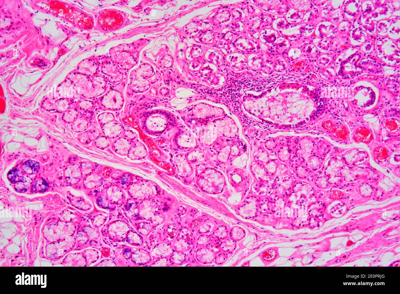
Sebaceous glands are simple acinar glands.

Label the photomicrograph of the sebaceous gland.. Skin Epidermis Dermis Subcutaneous Layer Human Thick Skin 25x At Dermis duct of sebaceous gland hair follicle sebaceous gland hair epidermis. Get more help from chegg. Learn vocabulary terms and more with flashcards games and other study tools.
This problem has been solved. Hair follicles sebaceous glands and sweat glands are all found in this region. Bottom layer epidermis piloerector muscle dermis hypodermis hair follicle.
Epidermis Sebaceous Gland Hair Follicle Duct Of Sebaceous Gland. Start studying Label the Skin. This problem has been solved.
Label the photomicrograph of compact bone. 30 Label The Photomicrograph Of. Labeling Parts of the Skin Identify the layers of skin.
This is a quiz called photomicrograph of thick skin and was created by member nhammond21. Oil glands that surround hair follicles. Allows parts of the skin to function and be controlled by the brain.
Label the photomicrograph of thick skin. Label the photomicrograph of thin skin quizlet. Label the structures of the hair follicle.
Secrete oils that lubricates skin hair and into the neck of the hair follicle. Erforoha ng statements as true t or false f. Show transcribed image text.
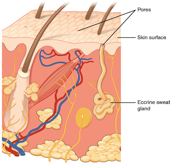
They are flask-shaped with short ducts that usually open into a hair follicle although some of them open directly onto the skin surface. Sebaceous glands produce an oily secretion called sebum. Learn vocabulary terms and more with flashcards games and other study tools.
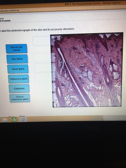
Label the photomicrograph of the skin and its accessory structures. Label The Photomicrograph Of The Sebaceous Gland. Written By Amber W Fox Sunday September 5 2021 Add Comment Edit Amber W Fox Sunday September 5 2021 Add Comment Edit.
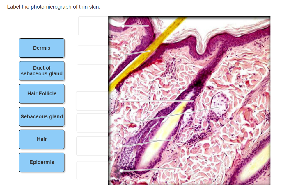
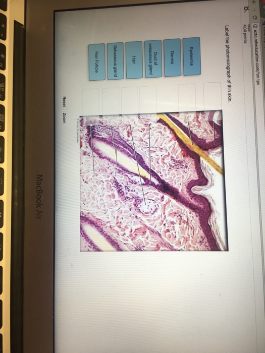
Show transcribed image text. Label the photomicrograph of thin skin. Epidermis Sebaceous Gland Hair Follicle Duct Of Sebaceous Gland Label The Photomicrograph Of The Skin And Its Accessory Structures.
Obtain a thin skin slide. Name the Layers of skin and label the dermal papilla and dermis Name the Layers of skin and label the dermal papilla and dermis. Name the layer of skin shown.
Anatomy And Physiology Support And Movement The. Label the photomicrograph of thin skin. Which structure is highlighted.
Start studying Figure 74 Photomicrograph of the skin and accessory structures. Then complete the statements that follow. Name the 4 layers of thin skin in both the cartoon and the photomicrograph.
Sebaceous gland Identify the following. Dermis stratum basale stratum spinosum stratum lucidum stratum corneum stratum granulosum basement membrane Identify the parts of this photomicrograph of skin. Play this quiz called photomicrograph of thin skin and show off your skills.
Which structure is highlighted. Written By Kim M Grant Friday August 13 2021 Add Comment. Epidermis hair follicle duct of sebaceous.
Before putting your slide on the microscope stage read the label inspect the slide with your eyes and make a note of any noticeable macroscopic features that may assist your inspection. 1 420 Skin Histology Stock Photos Pictures Amp Royalty Free. 35 Label The Photomicrograph Of The Sebaceous Gland.
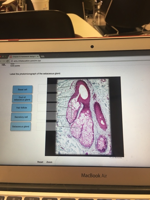
Label the photomicrograph of thin skin duct of sebaceous gland epidermis hair sebaceous gland dermis hair follicle. Label the photomicrograph of the sebaceous gland-Duct of sebaceous gland-Secretory cell-Sebaceous gland-Basal cell-Hair follicle-Hair follicle. Label the photomicrograph of the sebaceous gland.
Elongated cylinder orient runs through the core of each osteon. Your section might contain some glands whose section shows the continuity between the ducts lumen and the follicular space. Several sebaceous glands usually surround each hair follicle.
Label the photomicrograph of thick skin. The cartoon and the photomicrograph. Identify the layers of the epidermis with relation to their location and role in keratinization.
Chapter 12 Page 7 Histologyolm 4 0. Label the photomicrograph of the sebaceous gland. 33 Label The Photomicrograph Of Thin Skin.
Sebaceous gland piloerector muscle dermis hypodermis hair. Sections of thick skin or thin skin he apocrine sweat glands. With the 4X objective examine the slide.
Label the skin structures and areas indicated in the accompanying diagram of thin skin. Hair shaft epidermis dermal root sheath sebaceous gland dermis hair matrix. Learn vocabulary terms and more with flashcards games and other study tools.
There are usually 15 to 30 layers of cells in the stratum corneum. Expert Answer 100 22 ratings. Each gland has a single unbranched duct conveying secretory product from a cluster of flask-shaped secretory unit or acini.
Get more help from chegg. Play this quiz called photomicrograph of thin skin and show off your skills. Label the photomicrograph of the sebaceous gland.
Http Repository Uobabylon Edu Iq 2010 2011 5 2324 222 Pdf
 A P 1 Exercise 7 Activity 1 2 Ryk And Uyk Docx Lab Activity 1 Major Divisions Of The Skin Identify The Skin Structures In Figures 7 1 Course Hero
 Tubular Gland An Overview Sciencedirect Topics
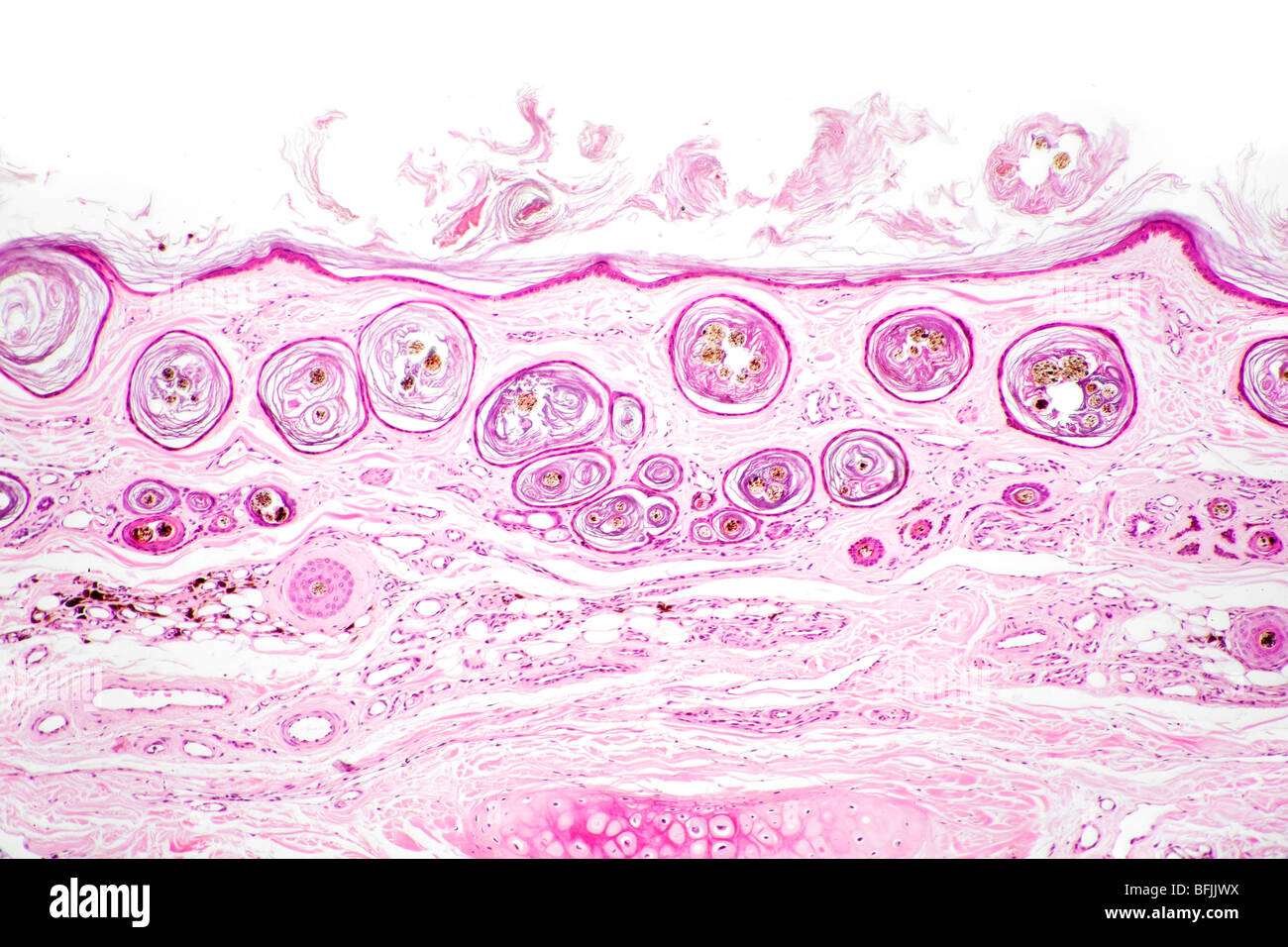 Brightfield Stained Photomicrograph Of Mammal Hair Follicles In Skin Cross Section Seen From Above Stock Photo Alamy

 A P Lab 3 Models Skin Hair Flashcards Quizlet
 Pdf Effects Of Microneedle Length Density Insertion Time And Multiple Applications On Human Skin Barrier Function Assessments By Transepidermal Water Loss
 Photomicrographs Showing Representative Labelling Of Various Skin Download Scientific Diagram
 Figure 7 4 Photomicrograph Of The Skin And Accessory Structures Diagram Quizlet

Chapter 12 Page 3 Histologyolm 4 0
:background_color(FFFFFF):format(jpeg)/images/library/3457/EZZZK6vnY0zfE0J1kUEEA_Capsule_of_Pacinian_corpuscle.png) Scalp And Hair Histology Kenhub
Scalp And Hair Histology Kenhub
Chapter 12 Page 8 Histologyolm 4 0
Histochemical Characterization Of Convict Cichlid Amatitlania Nigrofasciata Intestinal Goblet Cells
 Invasive Prostatic Adenocarcinoma Infiltrating Into Skeletal Muscle Download Scientific Diagram
Https Www Drcroes Com Uploads 8 1 2 8 8128660 Review Slides For Integumentary System Pdf
 Integumentary System Diagram To Label Lovely Human Skin Diagram Without Labels Integumentary System Free Graphic Organizers Printable Label Templates
 Anatomy Lab 4 Flashcards Quizlet
 Sebaceous Greeting Cards Fine Art America
 Skin 101 Hawaii Skin Cancer Coalition
 Photomicrograph Of A Section In The Skin Of An Albino Rat From Group P Download Scientific Diagram
 Label The Photomicrograph Of Thin Skin Dermis Duct Chegg Com
Https Www Msc Mu Com File Download Id 3601
Https Journals Sagepub Com Doi Pdf 10 1177 0192623319867322
 Label The Structures Of The Pelvis Pelvic Inlet Chegg Com
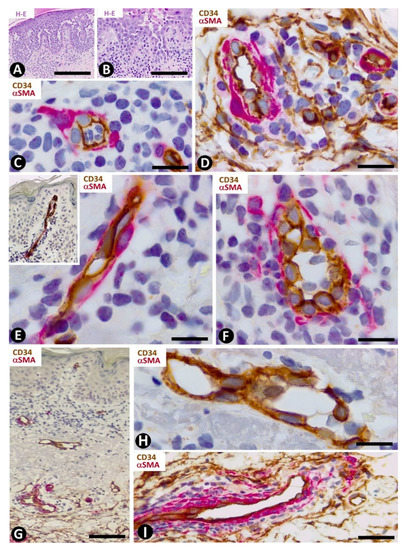 Ijms Free Full Text Cd34 Stromal Cells Telocytes In Normal And Pathological Skin Html
Https Nebula Wsimg Com 64f968542b4cb0a7d4d441f28eaf4933 Accesskeyid 2d26e20dc3b316743a3d Disposition 0 Alloworigin 1

 Label The Photomicrograph Of The Skin And Its Chegg Com
Moran Core Orbit Histopathology
Https Www Cell Com Stem Cell Reports Pdfextended S2213 6711 21 00040 0
 Accessory Structures Of The Skin Anatomy And Physiology I
Chapter 12 Page 8 Histologyolm 4 0
 Mucous Gland High Resolution Stock Photography And Images Alamy
 Integumentary System Histology Skin Labels Histology Slide Histology Slides Human Anatomy And Physiology Tissue Biology
 Cherenfant Ilab 4 Docx Bio251 Section 7 Week 4 Lab Roselaure Cherenfant Prof Sayed Figure 7 1 Diagram Of The Skin 1 2 3 4 5 Epidermis Papillary Layer Course Hero
 Power Point Lecture Slides Prepared By Barbara Heard
:watermark(/images/watermark_only.png,0,0,0):watermark(/images/logo_url.png,-10,-10,0):format(jpeg)/images/anatomy_term/stratified-cuboidal-epithelium/3iFoOuGxOtoBh1Rei0llA_Stratified_cuboidal_epithelium.png) Types Of Tissue Structure And Function Kenhub
Types Of Tissue Structure And Function Kenhub
 Skin And Body Membranes Ppt Download
:background_color(FFFFFF):format(jpeg)/images/library/3460/0y6tsYIMEudJBWtpAn8A_Hair_follicles_02.png) Scalp And Hair Histology Kenhub
Scalp And Hair Histology Kenhub
 Final Exam A P 1 Flashcards Quizlet

 Ch 6 Quiz Integumentary System Flashcards Quizlet
 Gastrointestinal Tract Sciencedirect
 31 Label The Photomicrograph Of Thin Skin Labels Design Ideas 2020
 Tubular Gland An Overview Sciencedirect Topics

 Label The Photomicrograph Of Thin Skin Quizlet
Https Www Drcroes Com Uploads 8 1 2 8 8128660 Review Slides For Integumentary System Pdf
Https Www Mdpi Com 2571 8800 4 3 23 Pdf
 Diagnosis And Management Of Gastrointestinal Neuroendocrine Neoplasms Surgical Pathology Clinics
 The Eye And Ocular Adnexa Chapter 47 Silverberg S Principles And Practice Of Surgical Pathology And Cytopathology

 Medium 10x Magnification Photomicrograph Of Thyroid Glands From X Download Scientific Diagram
 A P Lab 3 Models Skin Hair Flashcards Quizlet
 Skin 2 Accessory Structures Of The Skin And Their Functions Nursing Times
 Pages All Exocrine Glands Secretions Via Ducts Sebaceous Glands Sweat Glands Hair Hair Follicles Nails C 2015 Pearson Education Ppt Download
 A P Unit 2 Skin Tissue Model Photomicrographs Graphic Images Flashcards Quizlet
 Label These Structures Located In Axillary Skin Hair Chegg Com
 Integumentary System Diagram To Label Luxury Integumentary System Anatomy And Physiology Skin Anatomy Integumentary System Skin Anatomy Anatomy And Physiology
 Integumentary System Skin Anatomy Skin Model Integumentary System
Chapter 12 Page 3 Histologyolm 4 0
 Label The Photomicrograph Of The Skin And Its Chegg Com
 Sebaceous Greeting Cards Fine Art America

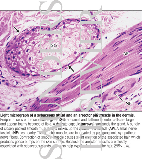 Light Micrograph Of A Sebaceous Gland And An Arrector Pili Muscle In The Dermis
 33 Label The Photomicrograph Of Thick Skin Labels Design Ideas 2020

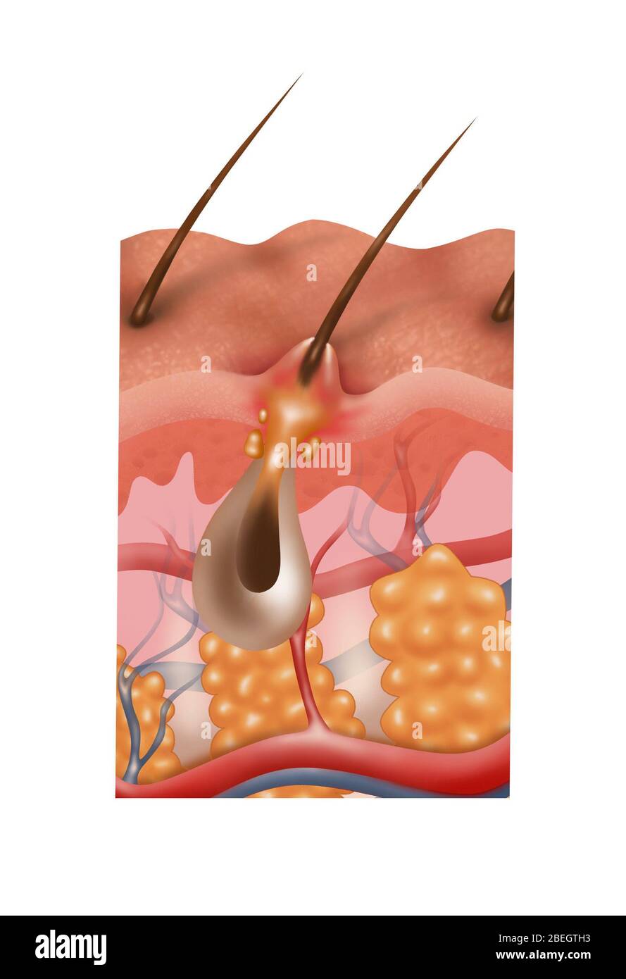 Hair Follicles Cross Section High Resolution Stock Photography And Images Alamy
 Lab 4 Integumentary System Flashcards Quizlet
Https Www Drcroes Com Uploads 8 1 2 8 8128660 Review Slides For Integumentary System Pdf
Chapter 12 Page 8 Histologyolm 4 0
 Male Reproductive System Sciencedirect
 Anatomy Lab Exam 3 Lab 9 Spinal Nerves Integument And Autonomics Flashcards Quizlet
 Sweat Gland High Res Stock Images Shutterstock
 Morphological Variants Of Meibomian Glands Correlation Of Meibography Features With Histopathology Findings British Journal Of Ophthalmology
Https Onlinelibrary Wiley Com Doi Pdf 10 1111 Brv 12579

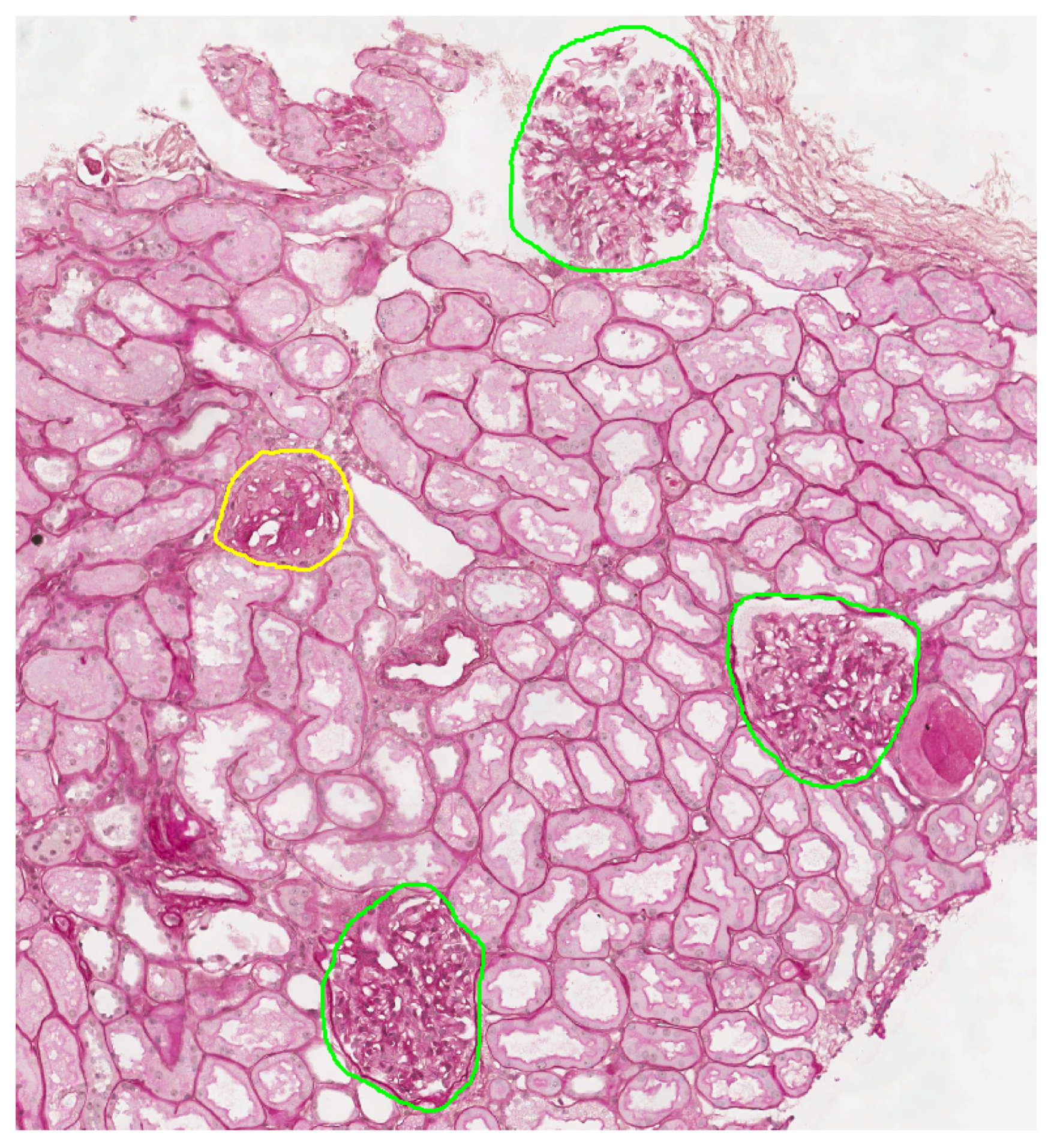 Electronics Free Full Text A Deep Learning Instance Segmentation Approach For Global Glomerulosclerosis Assessment In Donor Kidney Biopsies Html
 Morphological Variants Of Meibomian Glands Correlation Of Meibography Features With Histopathology Findings British Journal Of Ophthalmology
 Diagram Of Human Skin Structure Skin Structure Skin Anatomy Integumentary System
 Histopathologic Diagnosis Of Alopecia Clues And Pitfalls In The Follicular Microcosmos Diagnostic Histopathology
 Skin And Mammary Gland Sciencedirect
 Intralobular Duct An Overview Sciencedirect Topics

 Photomicrograph Of Skin Taken From The Same Animal And General Area As Download Scientific Diagram
Anatomy And Physiology Support And Movement The Integumentary System Viva Open
 Skin And Mammary Gland Sciencedirect

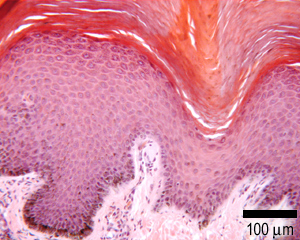

Epith_Skin_x63_001.jpg)